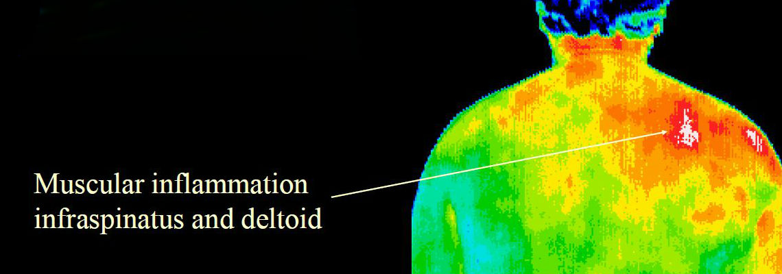
DITI can assist in the diagnosis, evaluation, monitoring and documentation of a large number of injuries and conditions, including soft tissue injuries and sensory/autonomic nerve fiber dysfunction.
Medical DITI can show a combined effect of the autonomic nervous system and the vascular system down to capillary dysfunctions. The effects of these changes show as asymmetries in temperature distribution on the surface of the body.


Clinical uses for DITI include:
– Defining the extent of a lesion of which a diagnosis has been previously made.
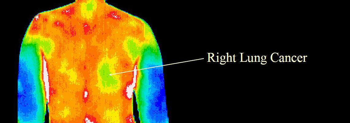
– Localizing an abnormal area not previously identified so that further diagnostic tests can be performed.
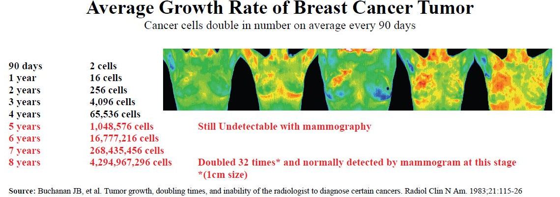
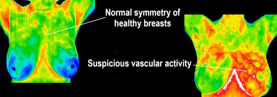
– Preventative Medicine-Detecting early lesions before they are clinically evident
– Monitoring the healing process before the patient returns to work or training.
Evaluating the extent of a known injury and the ability to monitor its healing process and response to treatment makes DITI an invaluable tool for physical therapists and health professionals involved in sports medicine.
Thermography is used as an aid for diagnosis and prognosis, as well as monitoring the effectiveness of a prescribed therapy or treatment for the following conditions and injuries:

| Back Injuries Arthritis Headache Nerve Damage Unexplained Pain Fibromyalgia RSD (CRPS) Dental & TMJ Artery Inflammation Vascular Disease |
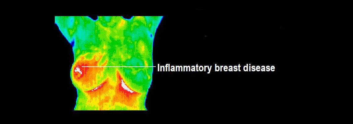
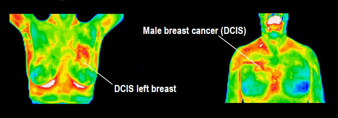
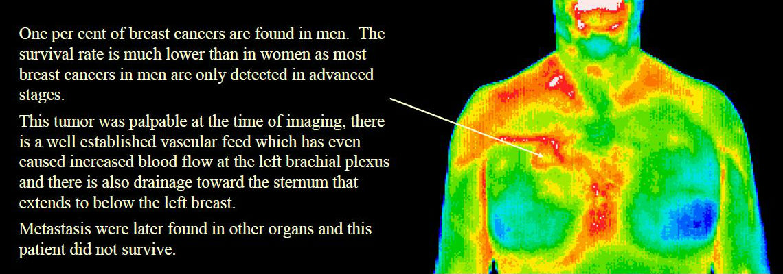
Breast Disease Carpal Tunnel Syndrome Disc Disease Inflammatory Pain Skin Cancer Referred Pain Syndrome Sprain/Strain Stroke Screening Whiplash Digestive Disorders |
Thermal Imaging in the Investigation of Deep Venous Thrombosis.
Preliminary assessment of clinically suspected deep venous thrombosis (DVT) of the lower limb by thermography avoids the need for over one third of venograms or duplex Doppler ultrasound scans. Clinical diagnosis of DVT is notoriously unreliable – hence the need for an accurate means of clinical investigation. Untreated DVT is dangerous as it can progress to pulmonary embolism (PE) which is frequently fatal or life-threatening. Treatment of DVT by anticoagulation poses risks of its own however, and should not be undertaken without a confirmed diagnosis. Thermal imaging is quick, simple, non- invasive, risk-free, cost-effective and highly sensitive in the initial investigation of suspected DVT; a negative thermogram excludes DVT and avoids the necessity for further investigation. Thermal imaging is, however, non-specific; a positive thermogram has a number of possible causes and is an indication for further assessment by venography or Doppler ultrasound to confirm or exclude DVT. Thermography should be considered the initial investigation of choice in clinically suspected DVT, proceeding to venography or Doppler ultrasound only when thermography is positive.